Olympus Multiphoton FVMPE-RS Apollo System
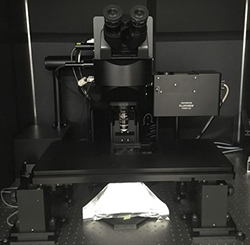 With the Olympus FVMPE-RS, countless possibilities for deep imaging in biological tissues to reveal both structural details and dynamic processes. The system delivers unmatched high-speed imaging, essential for capturing the dynamic in vivo response, with fine laser control pinpointing the precise site for optimum excitation efficiency — even deep within the sample. Accompanied by high S/N-ratio imaging and dedicated Olympus multiphoton objectives, efficient detection of scattered fluorescence photons is also ensured. In essence, the Olympus FLUOVIEW FVMPE-RS unites high-speed, deep observation capability with multi-color imaging and powerful laser light stimulation, offering a no compromise solution to answer innovative biological research questions. The upright microscope is coupled with a Spectra Physics Insight DeepSee laser with dual pulsed laser lines (1040nm, and 700-1300 tunable). The system contains a galvometer-based scanner for high-resolution imaging, a SIM scanner for uncaging and a resonant scanner for high-speed imaging applications.
Stellaris 8 Inverted Confocal/Multiphoton Microscope
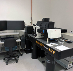 The Stellaris 8 inverted confocal system is ideal for imaging fixed or live cells and tissues. The system has a continuously adjustable white light laser covering excitation/emission from 440-790 nm along with a 405 diode laser. The system is equipped with 4 detectors (2 HydS and 2 HydX) and TauSense technology that allows the use of fluorescent lifetime-based imaging data to remove unwanted signal, measure changes in physiologic conditions, and separate overlapping spectra. Combining this with Fast Lifetime CONtrast (FALCON) allows for rapid kinetic studies in live cells, including FRET. The system also offers multiphoton capabilities with the Deep In Vivo Explorer (DIVE) to allow for imaging beyond a depth of 1mm in tissues and organs. The LAS X Navigator provides a quick overview of samples and allows for easy navigation to areas of interest.
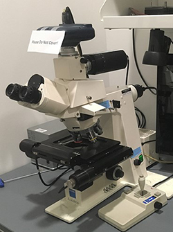
Zeiss AXIO Imager M2 Upright Widefield Fluorescent Microscope
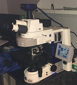 This is an upright fully motorized microscope, which includes a motorized stage that allows slide scanning (tiling) and acquisition of Z-stacks. The instrument is equipped with four filters that detect DAPI, GFP/488, Cy3 and Cy5 wavelengths. An Axiocam 105 digital color camera and a high-resolution Axiocam 506 digital monochrome camera are installed on the microscope, allowing acquisition of either color or monochrome images. Zen Image analysis software that includes deconvolution, colocalization and extended depth of focus is available on the instrument workstation and on a second workstation dedicated to image processing.
Zeiss Axio Scan.Z1 Slide Scanner
 This is an upright fully motorized microscope, which includes a motorized stage that allows slide scanning (tiling) and acquisition of Z-stacks. The instrument is equipped with four filters that detect DAPI, GFP/488, Cy3 and Cy5 wavelengths. An Axiocam 105 digital color camera and a high-resolution Axiocam 506 digital monochrome camera are installed on the microscope, allowing acquisition of either color or monochrome images. Zen Image analysis software that includes deconvolution, colocalization and extended depth of focus is available on the instrument workstation and on a second workstation dedicated to image processing.
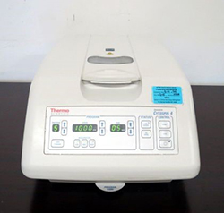
Leica CM195 Cryostat
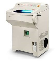 Available for tissue-thin sectioning. High specimen stability is achieved by adherence to deeply grooved specimen discs. Specimens freeze quickly because the pre-cooled discs feature a large back surface that fully contacts the freezing shelf with integrated Peltier element. UVC disinfection minimizes risk of contamination by infectious material. AgProtect, an antimicrobial, nanosilver coating on all outside surfaces also minimizes contamination risk.
NeuroLeucida and Stereo Investigator from MBF Bioscience
 Installed on a Zeiss Axio Skop upright microscope with a motorized stage and fluorescent capabilities, NeuroLeucida allows accurate reconstruction of cells permitting detailed morphometric analyses and 3D mapping. Stereo Investigator allows for unbiased estimates of the number, length, area and volume of cells or biological structures in a tissue specimen.
Cytospin Centrifuge
 Available to aid in affixing cell preparations to glass slides prior to imaging and is able to processes 12 specimens at one time. The cytocentrifuge deposits cells onto a clearly defined area of a glass slide and constructively flattens cells for excellent presentation and staining.
Keyence BZ-X800E
 An automated, all-in-one inverted digital microscope system equipped with 10, 20 and 40X objectives and filter cubes for detecting DAPI, GFP/488, Cy3 and Cy5 fluorochromes. The BZ-X800E includes powerful slide scanning capabilities that allow three slides at a time to be loaded for automated acquisition and stitching of bright field and/or fluorescence images. Stitching can also be combined with Z-stacking capability, which allows for capturing images within a range of focal planes that can then be projected as a single fully focused image. The instrument can accommodate a variety of sample formats including slides and standard tissue culture plates, dishes and flasks. The BZ-X800E comes equipped with an intuitive software platform that controls all objectives and filters. The included analysis software combines image correction tools with powerful quantification functions, including the haze reduction tool which instantly removes fluorescence blurring and enables visualization of weak fluorescence signals.
Dedicated Computer Workstations
Two workstations are available for post-acquisition processing of images. One workstation has Zen Pro image analysis software that includes deconvolution and colocalization packages. Another Workstation has Bitplane Imaris software (v. 8.02) for quantitation and 3D-rendering along with Neurolucida 360 for 3D reconstruction and analysis of neurons.