Research Seeks to Change Outcomes of Traumatic Brain Injuries

Researchers have advanced evidence for a treatment option that could prevent memory deficits and emotional problems in patients suffering from axonal injury caused by traumatic brain injuries (TBIs).
Axons are processes that connect a neuron to other neurons. These connections are crucial for every thought and feeling that we experience. When an axon is damaged, it cannot relay these messages to important processing centers in the brain.
Using a novel combination of high-resolution imaging and a GRIN lens implanted in the brain of mice, researchers documented axonal injury and the recovery process after a blunt injury to the brain. GRIN lenses are microscopic glass lenses that allows scientists to focus closely on the cells for good image quality. These lenses are smaller than Lincoln’s nose on a U.S. penny and were adopted by a handful of biologists to better study the brain.

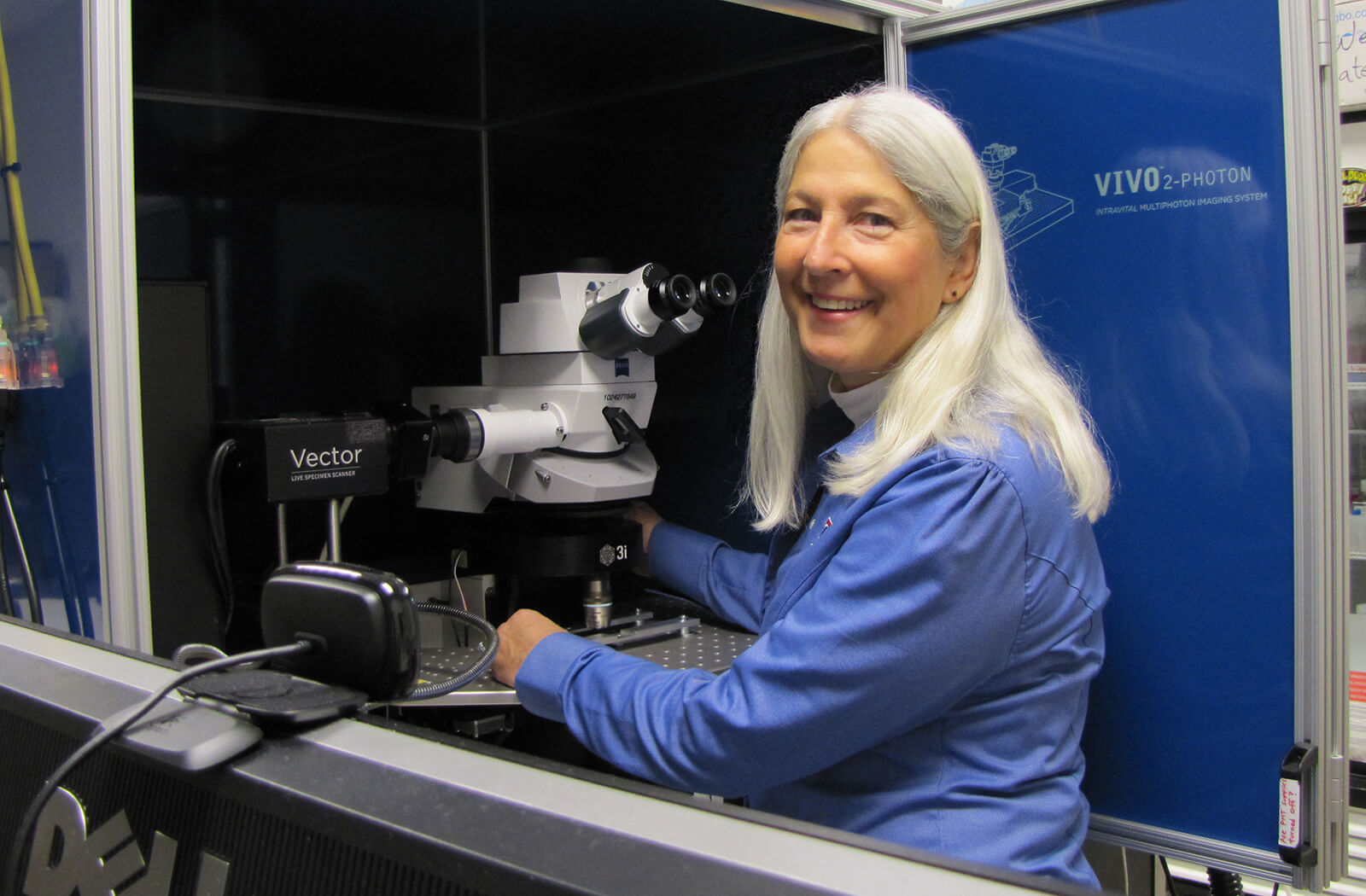
Jonathan Lifshitz, PhD, and Rachel Rowe, PhD, from the Translational Neurotrauma Research Program at the University of Arizona College of Medicine – Phoenix, collaborated with Chelsea Pernici, PhD, and Teresa Murray, PhD, from the Center for Biomedical Engineering and Rehabilitation Sciences at Louisiana Tech University. The study was led by Drs. Pernici and Murray who used a new imaging technology to show how diseases and injuries affect the brain over time and how medicines could reduce brain damage. This novel imaging was created in Murray's lab. Through this technology, the group of researchers were able to repeatedly image the same axons in the brain before and after brain injury, providing proof of TBI-related axonal pathology. Secondly, minocycline drug treatment promoted the recovery of injured axons.
“The fine communication elements of neurons — axons — are torn, ruptured and damaged, which contributes to a multitude of clinical symptoms,” said Dr. Lifshitz, director of the Translational Neurotrauma Research Program at the U of A College of Medicine – Phoenix. “To date, these processes have been inferred from fixed histological sections, clinical imaging and cells in culture. No one has observed an axon prior to injury, the consequence of injury and the outcome. Here, we show that axons do sustain injury and can either recover or become truncated.”

With treatment, a much larger percentage of the damaged axons healed, compared to those without treatment. As predicted, the immediate treatment resulted in the fewest number of damaged axons. Surprisingly, the delayed treatment resulted in the highest percentage of axons that healed from their injury. For both treatments (immediate and delayed), the damage did not spread as much to undamaged axons.
“Most brain injuries cause damage to just a few axons,” said Dr. Murray, an associate professor of Biomedical Engineering at Louisiana Tech University. “These few axons can’t be seen on MRIs and CT scans. So, most TBI patients are sent home with the hope that they will get better over time. However, even a few damaged axons can disrupt communication in the brain and affect neurological function.”
Dr. Murray developed an improved GRIN lens system to see more details of brain cells. Her team also designed 3D-printed hardware to help them find the exact same cells for each imaging session. Her improved GRIN lenses allowed the team to find and evaluate the same injured axons over several weeks post-injury.

No drug currently is on the market that is clinically used to prevent axon injury. The current results add evidence to support continued investigation of minocycline for TBI treatment.
“This study provides hope that we can use minocycline effectively if the drug can be taken early in the process and not given long-term. This could help reduce the inflammation, and thus some of the permanent damage caused by brain injury,” Dr. Murray said.
The team plans to continue their research by pairing minocycline with another drug to see if this early damage can be reduced further. If successful, this could greatly lessen the chances of memory loss and emotional problems for people suffering a TBI.
Topics
About the College
Founded in 2007, the University of Arizona College of Medicine – Phoenix inspires and trains exemplary physicians, scientists and leaders to advance its core missions in education, research, clinical care and service to communities across Arizona. The college’s strength lies in our collaborations and partnerships with clinical affiliates, community organizations and industry sponsors. With our primary affiliate, Banner Health, we are recognized as the premier academic medical center in Phoenix. As an anchor institution of the Phoenix Bioscience Core, the college is home to signature research programs in neurosciences, cardiopulmonary diseases, immunology, informatics and metabolism. These focus areas uniquely position us to drive biomedical research and bolster economic development in the region.
As an urban institution with strong roots in rural and tribal health, the college has graduated more than 1,000 physicians and matriculates 130 students each year. Greater than 60% of matriculating students are from Arizona and many continue training at our GME sponsored residency programs, ultimately pursuing local academic and community-based opportunities. While our traditional four-year program continues to thrive, we will launch our recently approved accelerated three-year medical student curriculum with exclusive focus on primary care. This program is designed to further enhance workforce retention needs across Arizona.
The college has embarked on our strategic plan for 2025 to 2030. Learn more.